By combining OCT and IVUS imaging, a more accurate image of the artery is achieved.
Infrared and Ultrasound sensors are placed next to each other on the catheter
OCTIVUS is inserted in the femoral artery and fed into the coronary artery through the aorta.
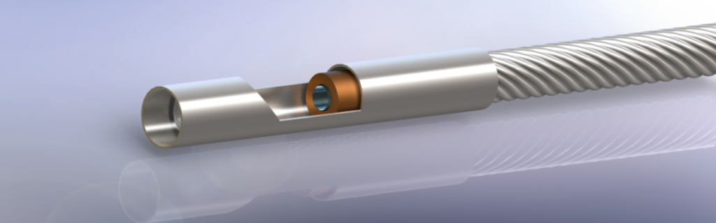
Figure 1. Construction of OCTIVUS
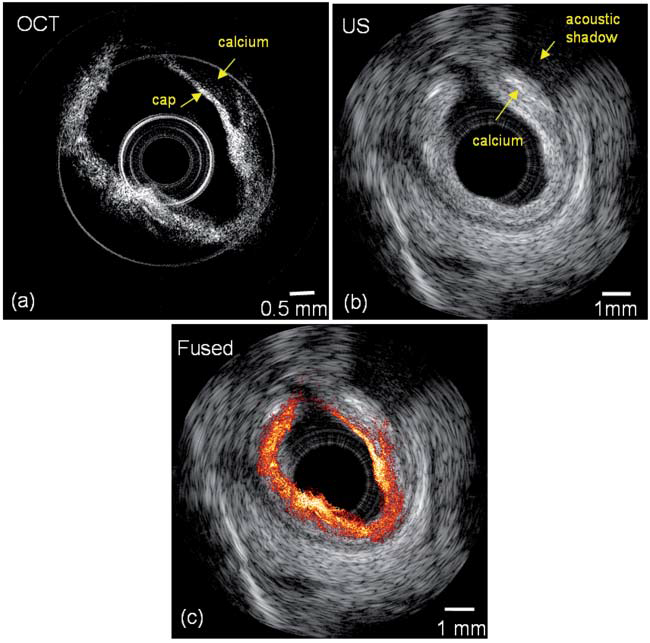
Figure 2. (a) Optical Coherence Tomography Image of Coronary Artery. (b) Ultrasound Image of Coronary Artery. (c) OCTIVUS Image of coronary artery.